THREE-DIMENSIONAL ULTRASOUND IN MILAN
AN EXAMINATION OF THE UTERUS WITH HIGH SPECIFICITY AND SENSITIVITY

Three-dimensional (3D) Ultrasound provides real-time realistic reconstruction of pelvic structures with three-dimensional images. It is the most recent technological evolution of Two-Dimensional (2D) Ultrasound.
It requires no previous preparation. Discomfort on the part of the patient is minimal, similar to normal Two-Dimensional Ultrasound. The best time for its performance is the second half of the menstrual cycle (in the advanced secretory phase), practically a few days before the arrival of menstruation. At this stage, the thicker, hyperechogenic endometrium better defines the contour of the endometrial cavity.
In particular, Three-Dimensional Ultrasound makes an accurate assessment of the fundus and internal conformation of the uterus. It provides detailed information on the external contour of the uterus with high specificity and sensitivity (100%). It allows very accurate detection of any abnormalities that need surgical correction. It evaluates and diagnoses cases of suspected uterine malformation ( septum, arcuate or bicornuate uterus etc.), myomas and uterine polyps that may be the cause of polyabortion, couple infertility and failure in assisted reproductive techniques. The regularity of the uterine cavity, in fact is a factor of absolute importance for the establishment and continuation of a pregnancy arising spontaneously or with assisted reproductive techniques. It plays, therefore, an important role in the study of couple infertility. It has replaced invasive methods such as hysteroscopy and laparoscopy.
Book a 3d ultrasound
OUR OFFICES
CHOOSE WHERE TO PERFORM THE ULTRASOUND
MILAN:
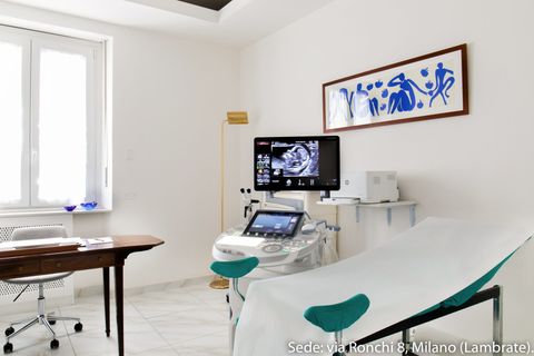
- - Studio Ginecologico Milano, in Via Ronchi 8, mezzanine floor, opposite train station exit and metro Lambrate, Rombon street side. Access for people with disabilities
MELEGNANO:
- Medical office located at 63 Castellini street Melegnano center staircase f, 9 floor, 3 elevators present, access for people with disabilities


